PsychNewsDaily Publishers
100 Summit Drive
Burlington, MA, 01803
Telephone: (320) 349-2484
PsychNewsDaily Publishers
100 Summit Drive
Burlington, MA, 01803
Telephone: (320) 349-2484
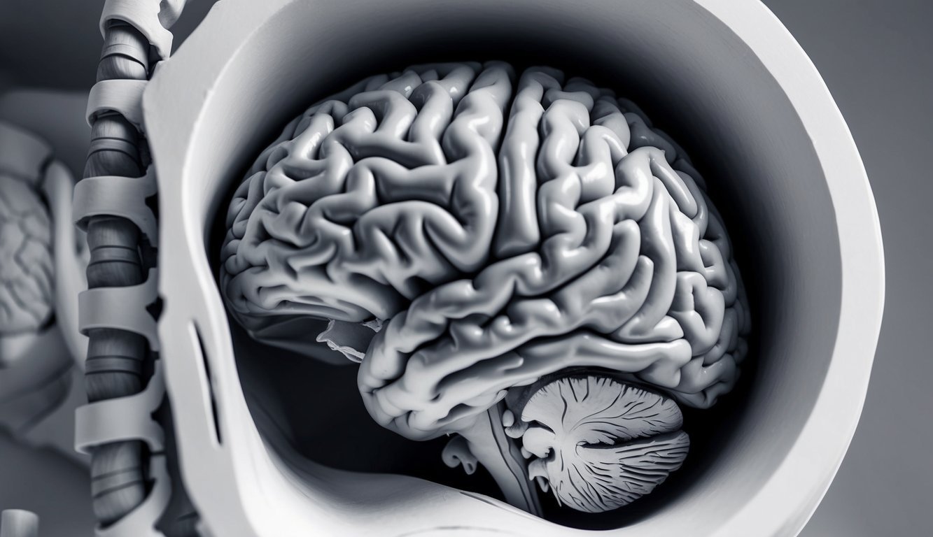
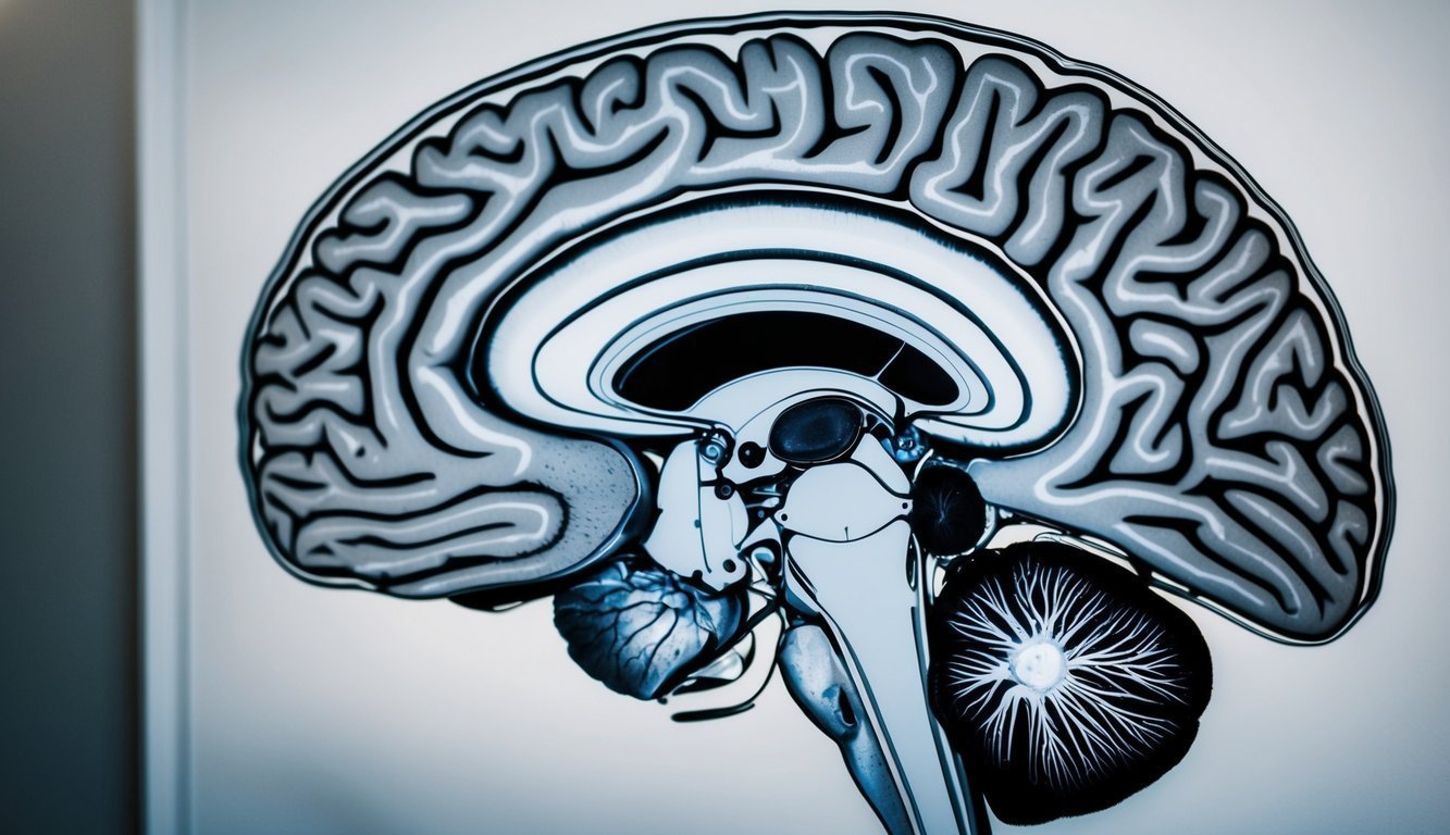
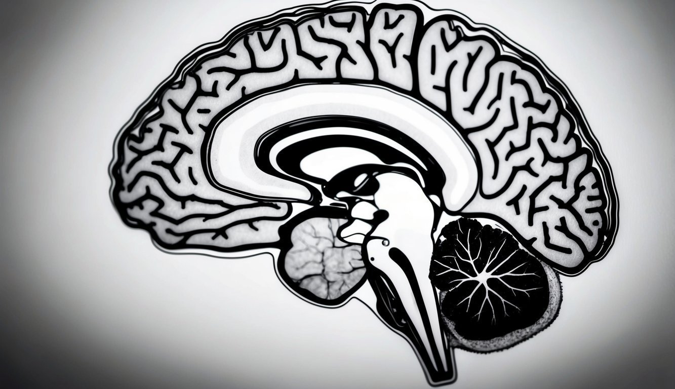
The human brain consists of the cerebrum, brainstem, and cerebellum, coordinating vital functions, sensory processing, and higher-order cognitive abilities through complex neural networks.

The human brain serves as a highly intricate organ at the heart of the central nervous system. A number of key structures collaborate to oversee bodily functions, process information, and produce thoughts and emotions.
The cerebrum, the brain’s largest part, is divided into two hemispheres and is crucial for higher-order functions such as reasoning, memory, and sensory processing. Its outer layer, known as the cerebral cortex, is composed of grey matter that includes neuron cell bodies.
Located beneath the cerebrum, the brainstem connects the brain to the spinal cord and regulates vital functions like breathing, heart rate, and blood pressure. The cerebellum, situated at the rear of the brain, is key for movement coordination and balance.
White matter consists of myelinated axons, establishing connections across different brain regions. The brain’s primary cell types, neurons and glial cells, play essential roles; neurons convey electrical signals, while glial cells offer support and protection.
Protective layers, known as meninges, encase the brain and spinal cord:
Cerebrospinal fluid circulates within and around these structures, providing cushioning and nutrients, as well as assisting in the removal of waste products from the brain.
A solid understanding of brain anatomy lays the groundwork for delving into psychological processes and their neural correlates.

The cerebral cortex is a sophisticated structure that plays an essential role in cognitive functions and behavior. Its complex organization into lobes and subcortical structures allows for diverse functions, from sensory processing to advanced thinking.
The cerebral cortex is comprised of four primary lobes, each responsible for unique functions. The frontal lobe governs executive functions, planning, and personality, and includes the motor cortex responsible for voluntary movements.
The parietal lobe engages in sensory information processing and spatial awareness via the somatosensory cortex. The temporal lobe facilitates auditory processing, memory formation, and language comprehension, housing Wernicke’s area, essential for understanding language.
Situated at the rear of the brain, the occipital lobe contains the primary visual cortex, critical for visual information processing. These lobes operate in unison, linked by intricate neuronal networks, to support advanced cognitive activities.
Beneath the cerebral cortex reside vital subcortical structures that assist various brain functions. The thalamus acts as a sensory and motor signal relay, while the hypothalamus is involved in regulating homeostasis and hormone production.
The basal ganglia are essential for motor control and learning. The limbic system, comprising the amygdala and hippocampus, plays a significant role in emotions, memory, and behavior. The amygdala processes emotional responses, and the hippocampus is crucial for forming new memories.
Known as the “master gland,” the pituitary gland oversees hormone production throughout the body. The pineal gland regulates sleep-wake cycles through melatonin production. These subcortical structures collaborate with the cerebral cortex to sustain cognitive functions and physiological processes.

The brainstem is integral to regulating essential bodily functions and autonomic processes. It operates as a relay center for information exchange between the brain and spinal cord, managing crucial involuntary functions vital for survival.
The brainstem is divided into three principal parts: the midbrain, pons, and medulla oblongata. The midbrain, positioned at the upper portion of the brainstem, is involved in visual and auditory processing, additionally aiding motor control and sleep regulation.
Located below the midbrain, the pons contributes to sleep, arousal, and respiratory regulation, containing nuclei essential for REM sleep and breathing rhythms.
The medulla oblongata, the brainstem’s lowest portion, is critical for sustaining vital functions. It governs:
Additionally, the medulla houses centers that control reflexes such as coughing, sneezing, and swallowing.
The brainstem is vital to the autonomic nervous system, which oversees involuntary bodily functions. It aids in the regulation of:
The brainstem’s function in autonomic control helps maintain homeostasis and respond to environmental changes. It processes sensory information from internal organs and modifies autonomic reactions accordingly.
Additionally, circadian rhythms that influence sleep-wake cycles and hormone release are partially managed by brainstem structures, aiding in the synchronization of various physiological processes with the external environment.

Neuroscience provides crucial understanding of the brain’s structure and functioning. This field investigates how diverse brain regions contribute to an array of cognitive processes and behaviors.
The cerebral hemispheres have specialized roles in brain function; typically, the left hemisphere is associated with language and logical reasoning, while the right hemisphere excels in spatial awareness and creativity.
Specific brain areas have been identified as responsible for motor functions and sensory data processing. The motor cortex governs voluntary actions, whereas the sensory cortex interprets incoming sensory information.
Crucial for higher-order thinking, decision-making, and planning, the prefrontal cortex is situated at the brain’s front. It also significantly influences personality and social behavior.
The formation and retrieval of memories require the cooperation of multiple brain regions. The hippocampus is essential for creating new memories, while other regions store long-term memories.
Neuroscientists explore how learned movements and imagination involve complex networks of neurons across various brain areas.
Recent research has examined the link between brain health and systemic issues like diabetes, highlighting potential new approaches to maintaining cognitive function.
Advancements in neuroimaging technologies have transformed our comprehension of brain structure and function, allowing researchers to observe the brain in action and offering unprecedented insights into human cognition and behavior.

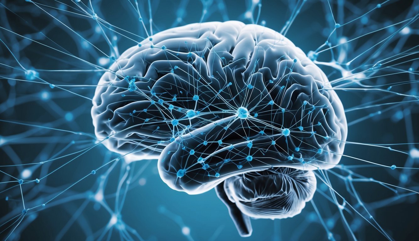
The extraordinary capabilities of the human brain arise from its detailed network of connectivity and communication. This intricate system is created by neurons, which transmit signals through synapses to develop neural pathways.
White matter, formed of myelinated axons, enables swift signal transmission between brain regions, while gray matter contains neuronal cell bodies responsible for processing information and generating responses.
The central nervous system, consisting of the brain and spinal cord, coordinates with the peripheral nervous system to govern bodily functions and movements. This complex network facilitates the development of motor skills and cognitive capabilities.
Functional connectivity in the brain refers to the temporal correlation of neural activity across different regions. This dynamic interaction adjusts to fulfill various tasks and respond to environmental stimuli.
In contrast, structural connectivity pertains to the physical neural pathways that connect brain areas. These structural links form the basis for functional interactions.
Key regions of the brain involved in connectivity include:
The Society for Neuroscience enhances our comprehension of brain connectivity and communication, delivering valuable insights for psychological and neuroscience research.